
Content
- What is lipoma in dogs
- Causes of lipoma in dogs
- Other Causes of Lipoma in Dogs
- Lipoma symptoms in dogs
- Diagnosis of lipoma in dogs
- Lipoma treatment in dogs

When we see that a dog has a lump, it can quickly come to mind that this is a tumoral process, something that alarms and worries tutors a lot when they think of the worst. It is true that on many occasions the tumors are malignant, but on many others they are also benign, the best example being the canine lipoma.
Lipoma in dogs is a tumor accumulation of fat cells or adipocytes. It is a benign tumor of mesenchymal origin that mainly affects older bitches of certain breeds, although no dog is free from suffering from it at any point in its life. Diagnosis is made using cytology, by observing a large number of adipocytes, and is usually not removed if it does not bother the dog and does not involve very deep layers of skin. Continue reading this PeritoAnimal article to learn more about lipoma in dogs - symptoms and treatment.
What is lipoma in dogs
A lipoma is a neoplasm or benign mesenchymal tumor which consists of the exaggerated accumulation of adipocytes, which are fat cells. It is a firm, soft and spongy tumor that can be solitary or multiple tumor nodules appear. Adipocytes are clustered with thin cell borders. When they are processed with methanol they dissolve into fat.
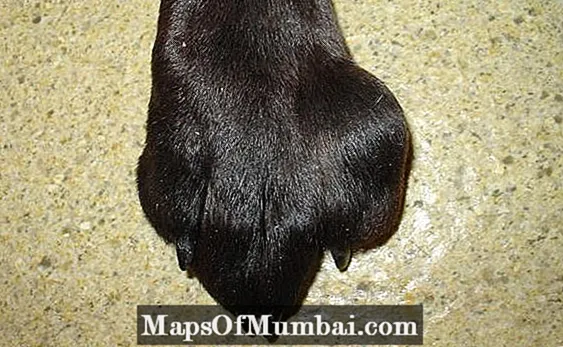
Lipoma in dogs develops in the subcutaneous tissue, especially of the limbs or the abdominal or thoracic cavity. Sometimes, cleaners can also include deeper layers, although not as common.
Perhaps you might be interested in this other article by PeritoAnimal in which we talk about cancer in dogs: types and symptoms.
Causes of lipoma in dogs
The main cause of lipoma in dogs is genetic character, with the most affected races being the following:
- Doberman.
- Cocker.
- Labrador retriever.
- German Shepherd.
- Pinschers.
It is usually more common in older dogs and females seem to be more susceptible. However, they can be detected at any age, race and gender.
Other Causes of Lipoma in Dogs
In addition to genetics, it is seen more often in dogs with overweight or obese, perhaps by a low-throughput metabolism that produces a low fat-metabolizing capacity, so that fat tends to accumulate.
They can also be caused by the body's inability to properly detoxify toxins by hepatic, intestinal or renal alteration.

Lipoma symptoms in dogs
Canine lipoma has a variable size, from less than 1 cm to several centimeters. If they are big they can pinch or annoy the animal, but in most cases it doesn't limit you in anything on a daily basis. Lipomas can be individual or appear several, and consist of consistency nodules:
- Firm.
- Soft.
- Soft.
- Encapsulated.
- Circumscribed.
- With sharp edges.
These tumors are usually located in the subcutaneous tissue of the limbs, neck, abdomen or chest. They tend to have good mobility as they usually do not bind to deep tissue, which is indicative of malignancy. However, they can sometimes grow in muscle tissue, appearing firmer, harder and less mobile, without indicating that they are malignant tumors.
THE evil variety Canine lipoma is liposarcoma, which can metastasize elsewhere in the dog's body, such as the bones, lungs or other organs. It is a lipoma-like but infiltrating tissue that invades muscle tissue and fascia. For more information, you can refer to this other article on dog tumors - types, symptoms and treatment.
Diagnosis of lipoma in dogs
The clinical diagnosis of cleanma in dogs is easy. After the detection of the nodule, it is considered a tumor process and one should go to the veterinary center to diagnose which type of tumor it is and whether it is benign or malignant. In the latter case, it should also be investigated for metastasis. The differential diagnosis of lipoma in dogs includes other canine nodules such as:
- Liposarcoma.
- Mast cell tumor.
- Soft tissue sarcoma.
- Sebaceous cyst.
- Epidermoid cyst.
- Histiocytoma.
The definitive diagnosis of lipoma in dogs is obtained with a Fine Needle Aspiration Puncture (PAAF), placing the cell content obtained on a slide and viewing it under a microscope, where a multitude of adipocytes will be observed, clarifying the diagnosis.
Adipocytes are seen as cells with vacuolated cytoplasm and a small, pyknotic, flat, and eccentric nucleus. If there is suspicion of involvement of deeper planes, it will be necessary advanced imaging tests, which will also help the surgeon plan the removal.
Lipoma treatment in dogs
The treatment of canine lipoma can be the surgical removal, but usually one chooses to leave it and observe its evolution. If it continues to grow to a considerable size, which causes discomfort, skin lesions or affects any structure in the dog, it should be removed.
Keep in mind that leaving a lipoma is not dangerous for your dog. These tumors do not metastasize or endanger the animal's life.
Now that you know all about lipoma in dogs, you might be interested in this video from our YouTube channel where we talk about the 10 dog breeds that live the longest.
This article is for information purposes only, at PeritoAnimal.com.br we are not able to prescribe veterinary treatments or perform any type of diagnosis. We suggest that you take your pet to the veterinarian in case it has any type of condition or discomfort.
If you want to read more articles similar to Lipoma in Dogs - Causes, Symptoms and Treatment, we recommend that you enter our Other health problems section.